NeuroMuscular Taping MEL Lines are fundamental to achieving precise, reproducible and clinically effective taping applications. By understanding the natural elasticity and biomechanical behaviour of the skin, clinicians can optimize decompression, enhance proprioceptive stimulation, improve lymphatic and vascular dynamics, and support evidence-informed rehabilitation through anatomically guided treatment strategies.
Understanding MEL Lines of Skin Elasticity
Successful NeuroMuscular Taping depends not only on selecting the correct treatment technique but also on understanding how the skin functions as a dynamic sensory and biomechanical interface. NeuroMuscular Taping MEL Lines (Major Elasticity Lines) provide clinicians with an anatomical framework for identifying the direction of maximum skin elasticity and functional movement, allowing tape applications to be tailored to the mechanical behaviour of each body region.
Furthermore, the effectiveness of NeuroMuscular Taping is influenced by multiple technical variables, including anatomical mapping, skin elasticity, tape width, coverage ratio and the principle of 0% tape tension. Rather than relying on compression, NeuroMuscular Taping applies elastic tape to pre-stretched skin to create tissue decompression, facilitating lymphatic drainage, improving microcirculation and enhancing neurosensory stimulation. By integrating MEL Lines with detailed clinical assessment and sound clinical reasoning, healthcare professionals can achieve more precise, reproducible and patient-specific rehabilitation outcomes across neurological, musculoskeletal, lymphatic and sports medicine settings.
The effectiveness of NMT is contingent upon a precise and anatomically informed application. As the technique relies primarily on cutaneous stimulation, its success depends on the clinician’s understanding of both skin function and underlying anatomical structures.
Skin directional elasticity variations define what I refer to as “lines of skin elasticity” (MEL – Major Elasticity Lines), which hold significant implications in both surgical planning and therapeutic interventions. Recognition and alignment with these lines are essential for optimizing tape application strategies, enhancing decompressive effects, promoting physiological tissue mobility, and minimizing unnecessary mechanical stress on the integumentary and fascial systems.. Official NMT courses provide the foundation necessary to safely and effectively apply this innovative method in neurodegenerative conditions. In this post, we break down the rehabilitation goals supported by the research and why proper certification is essential to translating theory into patient progress.
Why Skin Biomechanics is Important in NeuroMuscular Taping
The effectiveness of NMT is contingent upon a precise and anatomically informed application. As the technique relies primarily on cutaneous stimulation, its success depends on the clinician’s understanding of both skin function and underlying anatomical structures. The following core principles guide optimal taping outcomes:
- Anatomical and Functional Mapping Accurate identification of the affected structure(s), including muscle orientation, joint mechanics, and fascial lines.
- Skin Elasticity and Directionality Assessment of regional skin elasticity is essential to identify optimal application lines that correspond to the direction of maximum skin elongation or functional mobility restriction. A thorough understanding of skin biomechanics must be integrated with knowledge of the underlying musculoskeletal, vascular, neural, and lymphatic anatomy, as these structures directly influence the therapeutic objectives of the taping intervention.
- Zero Tension Application Tape must be applied without tension (0% tension) over previously stretched skin to produce the desired eccentric and decompressive effect.
- Tape Width Selection The width of the tape must correspond with the intended depth of action— narrower strips of tape for superficial drainage and wider strips for deeper muscular or joint effects.
- Coverage Ratio Consideration The ideal tape coverage is 30–50% of the skin surface in the treatment area. Excessive overlap or dense coverage may inadvertently create a compressive effect, counteracting the desired decompression and results expected.
These foundational principles not only ensure therapeutic precision but also maximise clinical efficacy, particularly in complex neurological, orthopedic and rheumatologic conditions where mechanical and neurovascular dysfunctions often coexist.
Skin Anatomy: Variability and Relevance in Taping
Anatomical and Functional Properties of the Skin
The skin is a highly dynamic and regionally variable organ, with its thickness, elasticity, and biomechanical properties influenced by numerous factors including gender, age, ethnicity, anatomical location, mechanical stress, and pathological conditions. Thickness typically ranges from 0.5 mm to 2 mm, with certain areas—such as the posterior neck, palms, and soles—reaching up to 4–5 mm. By contrast, the thinnest skin is found in regions such as the eyelids and external auditory canal.
Regional differences are consistent and clinically relevant:
- Flexor surfaces generally have thinner skin compared to extensor surfaces.
- Dorsolateral areas (e.g., head, neck, back) exhibit increased thickness.
- Ventral surfaces (e.g., abdomen, groin, and distal extremities) tend to be thinner.
- Maximum dermal thickness occurs in areas subject to mechanical load, such as the posterior cervical region, palms, and soles.
Elasticity and Biomechanical Orientation
Skin elasticity is another critical factor in therapeutic taping, especially in decompression methods such as NeuroMuscular Taping (NMT). Elastic properties vary significantly by location, depending on connective tissue composition and movement patterns. For example, a 3×100 mm skin segment from the lumbar or thigh region can stretch by 50% and support up to 10 kg of tension. Elasticity is generally greater in high-mobility areas—such as the anterior neck, mandibular region, and lateral trunk—while it is markedly reduced in more anatomically fixed zones like the nasal and auricular regions.
Chronic mechanical stress, pathological distension (e.g., overweight or fluid retention), or compromised vascularisation may result in the partial breakdown of dermal elastic fibers, clinically presenting as striae distensae (stretch marks) or age-related dermal atrophy.
Crucially, skin does not exhibit uniform elasticity in all directions. Its anisotropic mechanical properties result from the specific orientation and distribution of collagen and elastic fibers within the dermal layer. In the context of NeuroMuscular Taping and its eccentric decompression theory, these structural alignments are understood not merely as static skin tension lines, but as dynamic mobility vectors—reflecting functional lines of force that govern skin movement in response to underlying musculoskeletal activity.
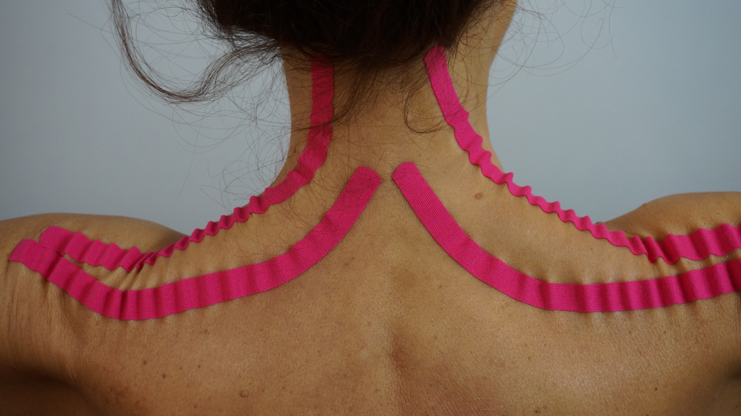
These directional variations define what I refer to as “lines of skin elasticity” (MEL – Major Elasticity Lines), which hold significant implications in both surgical planning and therapeutic interventions. Recognition and alignment with these lines are essential for optimizing tape application strategies, enhancing decompressive effects, promoting physiological tissue mobility, and minimizing unnecessary mechanical stress on the integumentary and fascial systems.
Skin Tension Lines: From Langer to Major Elasticity Lines (MEL)
The concept of “skin tension lines” originated in the 19th century with Karl Langer, who defined anatomical cleavage lines (Langer’s Lines) based on cadaveric puncture studies. These lines correspond to areas of lower skin elasticity and are widely used in surgical planning to guide incision orientation for optimal healing and scar minimisation. Later refinements, such as Kraissl’s Lines, were developed through in vivo studies and are particularly applicable to facial procedures due to their dynamic reference to muscular contractions.
In clinical practice and especially in the context of NeuroMuscular Taping, such biomechanical lines are of great significance. In 2000, the concept of Major Elasticity Lines (MEL) I introduced as a refinement of previous models. MELs represent functional trajectories of maximal skin elasticity and mobility, directly influenced by underlying muscular, fascial, and postural dynamics. In NMT, these lines serve as critical reference points for tape application to amplify the decompressive and proprioceptive effects of the technique (NeuroMuscular Taping Theory to practice. Blow David).
Comparative Diagram of Langer’s Lines, Kraissl’s Lines, and Major Elasticity Lines (MEL)
View. Blow, D (2012, 2018). NeuroMuscular Taping From Theory to Practice. ISBN: 9781467530361. David Blow’s book is a foundational text on the application of NeuroMuscular Taping (NMT) in rehabilitation. It offers an in-depth look at the theoretical principles underlying NMT, including its decompressive effects, facilitation of lymphatic drainage, and stimulation of mechanoreceptors for pain reduction. The book outlines for the first time the MEL lines of skin elasticity and REA range of elasticity fundamental to the NMT application methodology. Clinical examples and practical guidelines are outlined in the book for implementing NMT in the management of a range of conditions, including orthopedic, pediatric, neurology, lymphatic, rheumatological disorders. The integration of NMT with other rehabilitation strategies such as physiotherapy and manual therapy is also discussed, making it a comprehensive resource for clinicians and researchers interested in this therapeutic approach.
Functional Significance of Skin Folds and Elastic Mobility
Despite being anchored to deeper layers via the hypodermis, the skin maintains considerable freedom of movement. This mobility facilitates the formation of temporary or permanent folds that play important roles in proprioception and motor regulation. These folds often develop along joint axes or in response to repetitive muscle activity and serve as mechanical buffers that both facilitate and limit range of motion prior to the structural limits of muscles or tendons—acting as early warning systems for movement regulation.
- Temporary folds are induced by active muscle contraction and typically form perpendicular to the underlying muscle fiber orientation.
- Permanent folds result from long term mechanical stress, fascial adhesion, or scarring, and are commonly seen as anatomical landmarks with advancing age (e.g., horizontal cervical folds, periorbital lines, aging lines).
These structural features must be accounted for in clinical taping, particularly in decompression techniques. By aligning tape applications with MELs and respecting the natural folding patterns of the skin, NMT can enhance tissue mobility, reduce mechanical load on painful structures, and promote optimal sensorimotor integration.
Mapping MEL: Application Relevance in NeuroMuscular Taping
Effective application of NeuroMuscular Taping (NMT) hinges on the precise identification and alignment with Major Elasticity Lines (MEL)—functional trajectories of maximal skin elasticity defined through clinical observation and tactile feedback during movement and tape application. Unlike traditional anatomical references, which often underrepresent the biomechanical role of the integumentary system, MEL mapping reflects a dynamic, movement-oriented model of cutaneous and subcutaneous tissue behavior. Typically compression taping and bandage techniques cover Major Elasticity Lines at 90° or perpendicularly so as to enhance a reduction in functional trajectories and movement limitation. The primary therapeutic objective of aligning tape along MELs is to optimize decompression across multiple anatomical layers, from skin and fascia to muscle and vascular/lymphatic structures. Accurate MEL-based application facilitates:
- Restoration of local and regional mobility
- Resolution of interstitial congestion
- Modulation of cutaneous and subcutaneous mechanoreceptors
- Normalization of neurosensory and circulatory responses
In essence, MELs provide clinicians with practical landmarks for targeting decompressive effects, enhancing proprioceptive feedback, and supporting the body’s intrinsic regulatory systems.
The REA Concept: Range of Elastic Action
To complement MEL mapping, the Range of Elastic Action (REA) concept also introduced in 2000 to describe the angular flexibility and directional elasticity of skin in specific regions. Unlike MEL, which defines line-based tension trajectories, REA represents a functional “elastic window”—the angular arc (typically 0° to 45°) within which the skin can stretch and recoil in harmony with underlying structures.
REA values are derived from clinical observation of skin behavior during movement, and represent key factors in determining tape direction and orientation. Examples include:
- 0° REA: Found on the dorsal and flexor surfaces of metacarpophalangeal (MCP) and metatarsophalangeal (MTP) joints and indicates linear elasticity aligned exclusively along the longitudinal axis. Corresponds with single-plane muscle contractions (e.g., finger flexion-extension)
- 15° REA: Seen in the anterior knee region and allows limited angular mobility, consistent with hinge joint motion (flexion/extension)
- 25° REA: Observed around the wrist and permits multidirectional yet moderate movement, crucial for dynamic functional tasks
- 45° REA: Found in highly mobile areas such as the lateral and posterior cervical regions. Supports multi-planar movements including rotation, lateral flexion, and extension
Understanding REA helps clinicians determine the optimal angle of tape deployment, ensuring that elastic forces are distributed in accordance with the natural biomechanics of the skin-muscle-fascia continuum.
Clinical Reasoning for NeuroMuscular Taping Applications
To achieve reliable, reproducible, and patient-specific results using NMT, clinicians should:
- Identify MELs in the treatment area through palpation, observation of skin movement, and anatomical familiarity.
- Evaluate local REA to determine the most effective angular direction for tape application, respecting the elastic capacity of the region.
- Align tape with MEL trajectories to maximize cutaneous lifting and mechanical decompression.
- Avoid tape overlap that could result in unintentional compressive forces, potentially counteracting decompression goals.
- Respect natural tension vectors of the skin to enhance proprioceptive feedback and improve clinical efficacy.
This framework is particularly valuable in anatomically sensitive or biomechanically complex regions such as the cervical spine, lumbar region, and major joints (e.g., knees, shoulders), where skin mobility and tissue responsiveness play a significant role in both symptom expression and functional recovery.
Integrating MEL Lines into Evidence-Informed Rehabilitation
Are you interested in enrolling in a NeuroMuscular Taping (NMT) course? The NMT Institute offers internationally accredited certification programs across a wide range of clinical fields, including:
- Physical rehabilitation
- Neurology
- Oncology and post-surgical rehabilitation
- Sports therapy and athletic training
- Occupational and speech therapy
- Remedial education and nursing
- Vascular and lymphedema treatment and management
All NMT courses are delivered in-person in classroom settings, ensuring hands-on skill development, enhanced clinical reasoning, and optimal therapeutic outcomes. Online courses are not available, as practical application is central to the NMT therapeutic system.
To view upcoming course dates, detailed programs, and registration options, please contact your local course provider or visit the NMT Institute Website.
Planning to Include NMT in a Research Project?
A rigorously defined NeuroMuscular Taping methodology is essential for research transparency, reproducibility, and clinical impact. Whether you’re working on a clinical trial, thesis, or publication, a standardized approach enhances the validity, reliability, and translational value of your results.
To access the official NMT research methodology guidelines or request support in protocol development, contact us directly at: 📩 david.blow@nmtinstitute.org o info.taping@gmail.com

Are you interested in enrolling in an NMT Course?
The NeuroMuscular Taping Institute provides certification courses in physical rehabilitation, neurology, oncology, post surgical rehab, occupational therapy, speech therapy, remedial therapy, nursing as well vascular and lymphedema treatment and management. All trainings are classroom settings to enhance skill development and clinical reasoning creating optimum therapeutic results. ONLINE courses are not available. Please contact your local course provider or visit the NMT WEB SITE for course programs and dates.
For more details, course listings, and registration for an upcoming course CLICK HERE
Do you require validation of your NMT methodology for research purposes?
A detailed Neuromuscular Taping methodology is crucial in any research project for several reasons. Repeatability, Transparency, Validity and Reliability of your treatment choices will not only have an impact on your research but also an impact on future therapeutic choices. For more detailed information about the NMT, please contact us at: info.taping@mail.com
Know someone who might be interested in this newsletter? Share it with them.