Occipital Fascia NMT Treatment Protocol
The occipital fascia NMT treatment protocol is founded on a detailed understanding of the multilayered fascial architecture that connects the cranial base with the cervical spine, vascular structures and neural tissues. Rather than functioning as an isolated membrane, the occipital fascia forms a continuous biomechanical network that regulates pressure distribution, tissue mobility and neurovascular function. Through its decompression methodology, NeuroMuscular Taping (NMT) targets this strategic cranio-cervical interface to improve fascial mobility, optimize circulation and support functional recovery in neurological rehabilitation.
Introduction
The occipital fascia is one of the most complex connective tissue structures of the cranio-cervical region, integrating muscles, fascia, blood vessels, lymphatic pathways, peripheral nerves and the dura mater into a continuous functional system. The occipital fascia NMT treatment protocol recognizes that successful rehabilitation depends not only on anatomical knowledge but also on understanding how fascial architecture influences pressure regulation, movement, circulation and neuromuscular control.
Furthermore, the multilayered organization of the occipital fascia provides both structural stability and mechanical adaptability, allowing efficient transmission of forces between the skull and cervical spine. Restrictions caused by muscle hypertonicity, inflammation, scar formation or fascial densification may alter local biomechanics, reduce tissue mobility and compromise microcirculation and neurovascular function. By applying NeuroMuscular Taping according to decompression principles, clinicians can help restore physiological pressure gradients, improve vascular and lymphatic flow, enhance proprioceptive input and create favourable conditions for tissue healing and neuroplastic adaptation. This article examines the anatomical foundations and mechanical principles that support the occipital fascia as a key therapeutic target within NeuroMuscular Taping treatment protocols.Anatomical Fascial Architecture and Mechanical Role in NMT Treatment Protocol
The occipital fascia is a specialized component of the posterior cranio-cervical connective system located at the junction between the base of the skull and the upper cervical spine. Rather than functioning as an isolated membrane, it represents a continuum of layered fascial sheets integrating bone, muscle, vessels, nerves, and meninges.
From superficial to deep, the region includes:
- Superficial Fascial Layer • Continuous with the scalp fascia and subcutaneous tissues • Integrates cutaneous sensory innervation and superficial vessels • Interfaces with the posterior cervical fascial envelope
- Deep Muscular Fascial Layer • Encloses the trapezius, splenius capitis, semispinalis capitis, and longissimus capitis • Provides containment and sliding planes for muscular contraction • Maintains compartmental organization of posterior cervical tissues
- Suboccipital Fascial Compartment • Surrounds the rectus capitis posterior major/minor and obliquus capitis superior/inferior muscles • Anchors to the occipital bone periosteum and posterior atlanto-occipital membrane • Forms a dense myofascial cradle at the cranial base
- Myodural and Deep Connective Interfaces • Fascial continuities extend toward the spinal dura via myodural bridges • Integrates cranial periosteum, ligamentous structures, and dural envelopes • Mechanically links cranial motion with cervical spine dynamics
Fascial Layers and Cranio-Cervical Continuity
The occipital fascia forms a complex, multilayered connective tissue network that plays a fundamental role in integrating the anatomical and functional relationships between the cranial base and the cervical spine. Moreover, this fascial structure provides anchorage for the suboccipital musculature, stabilizing the small but highly specialized muscles responsible for fine head positioning and proprioceptive control. At the same time, it establishes a continuous structural link between the cranial periosteum and the deep cervical fascia, thereby connecting the skull to the broader fascial system of the neck and upper spine. Furthermore, the occipital fascia envelops the major neurovascular pathways emerging at the skull base, providing both mechanical protection and a dynamic sliding interface that allows these structures to adapt to movement and changing tissue tension.
As a result of these extensive anatomical connections, the occipital fascia functions as an essential conduit for the transmission and distribution of mechanical forces between the head and the cervical spine. Consequently, this fascial continuity ensures that movements of the cranium are efficiently coordinated with the underlying spinal structures, contributing to normal postural control and biomechanical efficiency. However, when fascial mobility becomes restricted by muscle hypertonicity, inflammation, connective tissue densification, or prolonged immobility, localized mechanical stress may propagate throughout the posterior kinetic chain. In turn, these restrictions can alter postural alignment, reduce cervical mobility, and disturb the coordinated relationship between the cranium and spine. Ultimately, these biomechanical disturbances may impair neuromuscular function, compromise tissue mechanics, and contribute to pain, functional limitations, and reduced movement efficiency.
The Mechanical Role of the Myodural Bridge
The cranio-cervical junction is a highly vascularized anatomical region where arterial inflow, venous return, and microvascular exchange occur within a confined yet mechanically dynamic environment. Importantly, unlike many peripheral regions of the body, circulation at the base of the skull is strongly influenced by local tissue pressure, fascial tension, and muscular tone. Consequently, even small variations in fascial compression or suboccipital muscle activity can influence vascular patency and the efficiency of blood flow. Furthermore, because this region integrates both cranial and cervical vascular pathways, it functions as a pressure-sensitive system in which mechanical forces and circulatory dynamics are closely interconnected. This unique relationship makes the cranio-cervical junction particularly responsive to therapeutic interventions aimed at restoring physiological tissue mechanics and fluid movement.
A central component of this circulatory system is the vertebral venous plexus, a network of valveless veins that surrounds the cervical spine and communicates directly with the intracranial venous sinuses. Unlike conventional venous systems, the absence of valves allows blood to flow bidirectionally according to pressure gradients between the cranial cavity and the spinal canal. While this anatomical arrangement provides remarkable adaptability for venous drainage under changing physiological conditions, it also makes the system particularly sensitive to external mechanical influences.
For example, increased fascial tension, muscular rigidity, connective tissue densification, or localized tissue compression may partially impede venous outflow, contributing to venous congestion and elevated interstitial pressure within the cranio-cervical tissues. As a result, impaired venous drainage may reduce microcirculatory efficiency, compromise tissue oxygenation, and alter the delicate balance between vascular, lymphatic, and interstitial fluid dynamics. Therefore, maintaining normal fascial mobility and physiological pressure gradients becomes an important objective in rehabilitation strategies designed to optimize circulation and support neurological function.
Pressure Regulation Within the Occipital Fascia
Arterial supply to the occipital region is provided primarily by occipital branches of the external carotid artery, together with contributions from vertebral and muscular arterial branches. These vessels supply the suboccipital muscles, deep cervical tissues, and adjacent neural structures. Efficient arterial inflow is necessary to maintain adequate oxygenation and metabolic support for the highly active neuromuscular tissues responsible for head positioning and postural control. However, arterial perfusion in this area can be indirectly influenced by surrounding tissue pressure, as excessive muscular contraction or fascial densification may increase extravascular resistance and reduce microvascular perfusion efficiency.
At the microscopic level, the region contains extensive microvascular networks, including capillaries and small arterioles that supply neural tissues, connective structures, and muscular fibers. These microcirculatory systems operate under delicate pressure balance, where capillary exchange of oxygen, nutrients, and metabolic by-products is governed by hydrostatic and osmotic gradients. Because capillary walls are highly sensitive to surrounding tissue pressure, elevations in interstitial pressure—such as those produced by muscular hypertonicity or fascial compression—can reduce capillary lumen diameter and limit effective perfusion.
Consequently, the cranio-cervical vascular environment is closely linked to the mechanical state of the surrounding tissues. When muscle tone is balanced and fascial structures maintain their physiological mobility, vascular pathways remain unobstructed and microcirculatory exchange proceeds efficiently. Conversely, increased tissue density or reduced fascial glide can alter pressure gradients within the vascular network, potentially compromising venous drainage, capillary perfusion, and metabolic exchange. For this reason, therapeutic approaches that reduce local mechanical pressure and restore tissue mobility—such as decompressive strategies used in NeuroMuscular Taping—may contribute to improved vascular dynamics and enhanced physiological regulation within the cranio-cervical region. Unlike peripheral circulation, venous return in this region is highly pressure-dependent. Fascial compression may:
- Reduce venous outflow efficiency
- Increase local vascular resistance
- Contribute to tissue hypoxia
- Promote inflammatory accumulation
Restoring fascial mobility improves microvascular perfusion and supports metabolic exchange in neurologically vulnerable tissues.
Local Blood Circulation and Fascial Mobility
Recent advances in anatomical and neurophysiological research have highlighted the fundamental role of lymphatic circulation in maintaining the physiological homeostasis of the central nervous system and surrounding cranial tissues. The discovery and characterization of meningeal lymphatic vessels have demonstrated that the brain and its protective membranes possess specialized drainage pathways that contribute to the removal of metabolic by-products, excess interstitial fluid, and immune mediators. Within this context, the occipital and cranio-cervical region represents a critical anatomical gateway through which cranial lymphatic and interstitial fluids transition toward the cervical lymphatic system.
The posterior cranial base, including the occipital region, functions as a convergence zone where superficial lymphatic vessels of the scalp and posterior neck meet deeper lymphatic channels associated with the cervical fascia and musculature. These superficial lymphatics collect fluid from the integumentary tissues of the scalp and surrounding connective structures, directing it toward posterior auricular and occipital lymph nodes. From these nodes, lymphatic flow continues toward the upper deep cervical lymphatic chain, which serves as the principal drainage route for fluids exiting the cranial and cervical compartments.
Beneath these superficial structures, deep cervical lymphatic collectors receive drainage from intracranial and meningeal lymphatic vessels that accompany dural venous sinuses and cranial nerve pathways. These deep collectors ultimately channel lymph toward the internal jugular lymphatic trunks, which represent one of the primary exit routes for cranial lymphatic drainage. Because of this anatomical arrangement, the cranio-cervical junction acts as a transitional interface where intracranial and extracranial lymphatic systems integrate and coordinate fluid clearance from the head and neck. An additional component of this system involves the regulation of interstitial fluid movement at the cranial base. Interstitial fluids originating from neural tissues, connective structures, and vascular filtration processes must be efficiently transported away from the cranial cavity to maintain stable pressure and biochemical conditions. The occipital region provides one of the principal pathways through which these fluids exit the cranial environment and enter the cervical lymphatic circulation. Effective drainage depends on the maintenance of appropriate pressure gradients between cranial tissues, lymphatic vessels, and surrounding fascial compartments.
Because lymphatic vessels operate as low-pressure transport systems, their function is particularly sensitive to the mechanical state of the surrounding tissues. Increased muscular tension, fascial densification, or external compression in the occipital and cervical region can alter the pressure gradients necessary for efficient lymphatic uptake and flow. Conversely, when fascial mobility is preserved and tissue pressure remains balanced, lymphatic channels can function more effectively, supporting fluid clearance and contributing to the regulation of local inflammatory and metabolic processes. For this reason, therapeutic strategies that promote decompression and improve fascial mobility at the cranio-cervical junction may indirectly support lymphatic drainage from the cranial base. By facilitating the movement of interstitial fluids toward cervical collectors, such interventions help maintain optimal fluid balance and may contribute to improved neurological and musculoskeletal function within the head and neck region.
Fascial restriction may impair fluid transport, contributing to:
- Interstitial congestion
- Increased tissue pressure
- Delayed inflammatory resolution
- Impaired neural recovery environments
Optimizing fascial glide facilitates fluid dynamics essential for neurological recovery.
Local Pressure Regulation and Neural Sensitivity
The suboccipital region represents one of the most neurologically specialized areas of the musculoskeletal system. Within the limited anatomical space between the occipital bone and the upper cervical vertebrae lies a dense network of neuromuscular and connective structures responsible for fine control of head position, postural regulation, and sensory feedback to the central nervous system. The suboccipital space contains an exceptionally high concentration of proprioceptive receptors, mechanosensitive nociceptors, and autonomic neural interfaces, all embedded within the surrounding fascial and muscular tissues.
The small suboccipital muscles—such as the rectus capitis posterior major and minor, and the obliquus capitis superior and inferior—contain a particularly high density of muscle spindles, specialized proprioceptive receptors that detect subtle changes in muscle length and tension. These receptors provide continuous feedback regarding head orientation and movement, allowing the central nervous system to regulate balance, gaze stabilization, and postural alignment. Through their connections with vestibular and visual control systems, these proprioceptive pathways play a critical role in coordinating head–neck positioning with overall body posture.
In addition to proprioceptive structures, the suboccipital region contains numerous mechanosensitive nociceptors located within the fascia, muscular tissues, and surrounding connective structures. These sensory receptors respond to mechanical deformation, compression, or tension within the tissues. When fascial tension increases or when muscular hypertonicity produces sustained compression, these nociceptors may become sensitized, contributing to local pain perception and potentially facilitating cervicogenic headache patterns. Increased mechanical loading can therefore alter nociceptive signaling pathways and influence both local discomfort and broader neural responses.
The cranio-cervical junction also contains important autonomic neural interfaces, including sympathetic fibers that regulate vascular tone and local microcirculation. These neural elements contribute to the modulation of blood flow and tissue metabolism within the head and neck region. Because autonomic fibers are sensitive to mechanical stress, excessive fascial tension or compression within the suboccipital compartment may disrupt their regulatory function, potentially influencing vascular dynamics and neuromuscular tone.
Elevated fascial tension within the occipital region can therefore produce a cascade of neurophysiological effects. Increased mechanical pressure on proprioceptors may distort afferent sensory input, altering the brain’s interpretation of head position and movement. At the same time, compression of mechanosensitive nociceptors may amplify pain signaling pathways, while disruption of autonomic interfaces may affect vascular regulation and muscle tone. These changes can interfere with the integration of postural reflexes and sensorimotor coordination, ultimately contributing to inefficient movement patterns and reduced postural stability.
Conversely, when local tissue pressure is reduced and fascial mobility is restored, the neural environment of the suboccipital region can return toward physiological balance. Decompression of the fascial layers allows proprioceptive receptors to function within their normal mechanical range, supporting more accurate sensory feedback and improved coordination between cervical musculature, vestibular inputs, and visual orientation systems. At the same time, reduced mechanical stress on nociceptive structures may decrease pain signaling and normalize muscle tone regulation. Through these mechanisms, optimization of local pressure conditions enhances neurosensory modulation, improves postural reflex integration, and supports more efficient functional control of head and neck movement. Elevated fascial tension increases mechanical pressure on these structures, potentially altering:
- Pain signaling pathways
- Muscle tone regulation
- Postural reflex integration
- Sensorimotor coordination
Reducing local pressure enhances neurosensory modulation and functional control.
NeuroMuscular Taping and Fascial Decompression
Due to its strategic role in regulating cranio-cervical biomechanics, vascular dynamics, and neural integration, the occipital fascia represents a key therapeutic target in NeuroMuscular Taping (NMT)–based neurorehabilitation. Its position at the interface between the cranium and cervical spine allows local mechanical and fluid changes to influence broader neurological function. As a result, decompressive interventions at the occipital level can play a significant role across a wide spectrum of central and spinal neurological disorders.
- In stroke rehabilitation, occipital NMT applications support cranial venous return and help reduce secondary interstitial congestion, creating a more favorable environment for neural recovery. By improving sensory input from the cranio-cervical region, these interventions assist in sensorimotor reintegration and contribute to improved head–neck postural control, which is essential for functional recovery and balance.
- In Parkinson’s disease, where axial rigidity and altered postural reflexes are prominent, occipital decompression helps reduce cervico-cranial compression and modulate abnormal muscle tone patterns. By improving tissue compliance and reducing mechanical resistance, NMT may enhance tolerance to upright posture and facilitate more efficient postural adjustments.
- In individuals with cerebral palsy, characterized by persistent abnormal tone and impaired motor control, occipital treatment contributes to modulation of extensor hyperactivity and supports improved cranio-cervical alignment. This facilitates neuromotor training by creating a more adaptable mechanical environment, while also improving comfort and positioning during both therapeutic activities and daily care.
Beyond these conditions, occipital NMT interventions are relevant in a range of central and spinal neurological pathologies, including traumatic brain injury, spinal cord injury, multiple sclerosis, neurodegenerative disorders, and chronic central pain syndromes. Across these conditions, dysfunction in the cranio-cervical region often contributes to altered pressure regulation, impaired fluid dynamics, and disrupted neuromuscular coordination. The primary therapeutic objectives of occipital NMT applications include normalization of local tissue pressure, optimization of vascular and lymphatic fluid exchange, modulation of neuromuscular activity, and support of postural re-education. By creating a decompressive environment within the occipital and upper cervical region, NMT enhances the physiological conditions necessary for effective rehabilitation.
From a clinical perspective, targeted occipital intervention contributes to improved local hemodynamics, facilitating more efficient blood flow and oxygen delivery. Enhanced lymphatic clearance supports the removal of interstitial fluid and inflammatory mediators, while reduction of mechanical stress on nociceptive structures decreases pain and neural irritability. In parallel, improved fascial mobility and muscle coordination lead to more efficient cervical biomechanics and better integration of head–neck movement within global postural strategies.
In neurorehabilitation, relatively small anatomical regions can exert profound system-wide effects. The occipital fascia is one such region, where precise and sustained intervention through NeuroMuscular Taping can influence circulation, neural regulation, and overall functional recovery.
Clinical Implications for NMT Treatment Protocols
Integrated Treatment of Occipital Rigidity: Occipital rigidity is rarely an isolated local dysfunction. In NeuroMuscular Taping, it is addressed through a regional-to-global decompressive strategy that restores tissue mobility, reduces neuromuscular hypertonicity, and improves fluid dynamics across the cranio-cervico-thoraco-diaphragmatic continuum. This series of treatment protocols follows NMT decompression principles: skin displacement → zero tape tension → visible convolutions → space creation → pressure normalization → activation
Clinical Objectives: • Reduce suboccipital hypertonicity • Restore cranio-cervical mobility • Decompress neurovascular interfaces • Improve venous and lymphatic outflow • Normalize postural load distribution • Support neurorehabilitation biomechanics
NMT Applications: Treatment Sequence (Proximal → Distal Integration)
1️⃣ Occipital Application — Primary Decompression
- Target: Occipital fascia & suboccipital compartment Goal: Reduce local pressure, improve cranial drainage
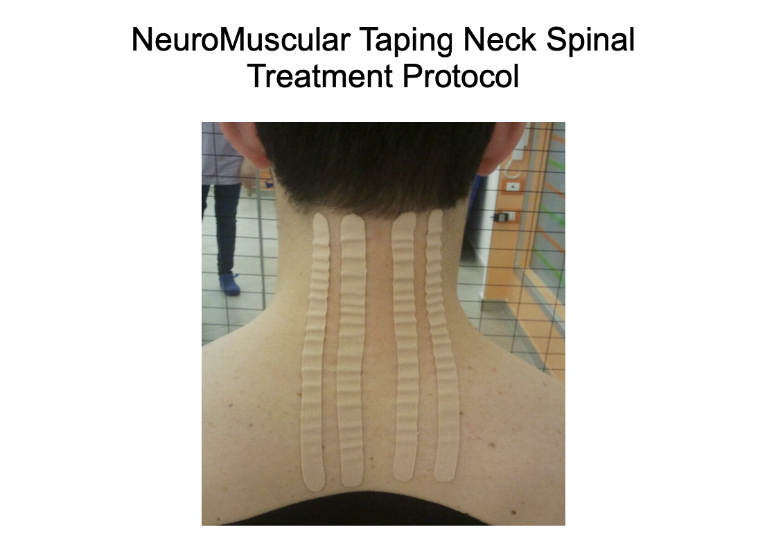
- Technique • Patient seated, cervical flexion • Manual cranial skin displacement • C2 → T4 orientation • Central and bilateral decompressive I-strips • Zero stretch, visible convolutions
- Clinical Effect → Cranio-cervical decompression → Reduced occipital density → Improved efficiency of occipital blood flow → Improved venous outflow
2️⃣ Neck Spinal Application — Cervical Axis Release
- Target: Deep cervical fascia & paraspinal chain Goal: Reduce axial compressive load
- Technique • Skin displaced laterally • Longitudinal decompressive strips along cervical paraspinals • C2 → T4 orientation • No tension
- Clinical Effect → Reduced segmental compression → Improved cervical glide → Neural interface unloading
3️⃣ Rhomboid Application — Posterior Stabilizer Balance
- Target: Rhomboid fascia & scapulo-thoracic interface Goal: Normalize posterior chain tension
- Technique • Scapula in max. protraction • Horizontal decompressive strips thoracic spine → medial scapular border
- Clinical Effect → Reduced dorsal fascial pull → Improved scapular mechanics → Indirect cervical unloading
4️⃣ Sternocleidomastoideo Application — Anterior–Lateral Balance
- Target: SCM fascial envelope Goal: Reduce rotational strain on cranio-cervical junction
- Technique • Head rotated contralaterally • Skin gently displaced • Decompressive strip mastoid → sternoclavicular region (2 strips)
- Clinical Effect → Reduced anterior-lateral tension → Improved head positioning → Reduced cervical tension → Vestibulo-cervical support
5️⃣ Anterior Neck Fascia Fascial Glide
- Target: Infrahyoid & pretracheal fascia Goal: Restore anterior cervical sliding planes
- Technique • Head and Neck extended • Longitudinal decompressive strips along the anterior fascia • Gentle longitudinal skin displacement
- Clinical Effect → Reduced anterior fascial tightness → Improved swallowing–breathing synergy → Cervical mobility support
6️⃣ Scalenius Muscle Group — Neurovascular Gateway
- Target: Deep lateral cervical compartment Goal: Reduce brachial plexus & vascular compression
- Technique • Head slightly side-flexed • Vertical decompressive strips lateral cervical column
- Clinical Effect → Thoracic outlet decompression → Improved neck posture → Improved upper limb neurodynamics → Reduced cervical guarding
7️⃣ Superior Trapezius — Load Redistribution
- Target: Upper trapezius fascial sheath Goal: Reduce cranial traction forces
- Technique • Shoulder relaxed and head contra-laterally flexed • Oblique strips occipital region and C5 → acromion • Tissue displacement before application
- Clinical Effect → Reduced cranial drag → Improved shoulder–neck coordination → Postural load balance
8️⃣ Abdominal Application — Core Pressure Regulation
- Target: Abdominal fascial cylinder Goal: Normalize intra-abdominal pressure transmission
- Technique • Supine position • Decompressive longitudinal abdominal strips • Encourage fascial expansion
- Clinical Effect → Improved pressure distribution → Reduced axial load transfer → Postural stabilization support
9️⃣ Anterior Functional Diaphragm — Pressure System Reset
- Target: Respiratory diaphragm fascial interface Goal: Optimize cranio-caudal pressure gradients
- Technique • Supine position with the arms over the head. Costal margin decompressive fan strips • Gentle downward skin displacement
- Clinical Effect → Improved respiratory mechanics → Enhanced venous–lymphatic return → Global pressure regulation
Conclusion
The restoration of functional cranio-cervical mobility requires an integrated therapeutic approach that extends beyond isolated local treatment. In NeuroMuscular Taping, optimal outcomes are achieved through the coordinated interaction of multiple biomechanical and physiological systems. Effective occipital decompression must be combined with scapulo-thoracic balance, anterior fascial glide, and respiratory pressure normalization to create a stable and adaptable cranio-cervical environment. This systemic integration allows mechanical loads to be distributed efficiently, pressure gradients to be regulated, and fluid dynamics to function in a physiologically optimal manner.
When these components are addressed collectively, the clinical outcomes are both measurable and functionally meaningful. Patients typically demonstrate a reduction in occipital hypertonicity, improved head–neck mobility, and greater postural endurance. Decreased cervicogenic pain reflects reduced nociceptive input and improved tissue compliance, while enhanced neuromuscular coordination supports more efficient movement patterns. Importantly, these changes contribute to improved readiness for neurological rehabilitation, creating the conditions necessary for more effective motor relearning and functional recovery. In this context, the occipital region serves not only as a local treatment site but as a key regulatory interface influencing global postural and neurological function.
References
Yahia LH, Rhalmi S, Newman N, Isler M. Sensory innervation of human thoracolumbar fascia. Spine (Phila Pa 1976).1992;17(11):1313–1317. https://doi.org/10.1097/00007632-199211000-00006
Andi WS, Wahida W, Blow D. Application of neuromuscular taping on occipital neuralgia: Case study. October 2019. Journal of the Neurological Sciences 405:46-47.https://www.sciencedirect.com/science/article/pii/S0022510X19321148
Blow D. NeuroMuscular Taping: From Theory to Practice. (2012). ISBN 10: 1467530360 / ISBN 13: 9781467530361
Blow D. Neuromuscular Taping: Treatment of Edemas, Hematoma. (2013) ISBN 10: 8870515990 / ISBN 13: 9788870515992
Costantino C, Pedrini MF, Licari O. Neuromuscular taping versus sham therapy on muscular strength and motor performance in multiple sclerosis patients. Disabil Rehabil. 2016;38(3):277–281. https://doi.org/10.3109/09638288.2015.1038365
Findley TW. Fascia research from a clinician/scientist’s perspective. Int J Ther Massage Bodywork. 2009;2(4):1–6. https://doi.org/10.3822/ijtmb.v2i4.63
Kristianto H, Ahsan A, Mukaroma L, Akhsinnadya F, Indri MC, Damayanti N, Kusbandiyah I, Rohyani RT, Blow D, Rias YA, Wibisono AH, Noviyanti LW, Nasution TH. Effects of neuromuscular taping form I on wound temperature and erythema in diabetic foot ulcer: a preliminary study. J Wound Care. 2024;33(6):425–430. https://doi.org/10.12968/jowc.2020.0291
Levick JR, Michel CC. Microvascular fluid exchange and the revised Starling principle. Cardiovasc Res. 2010;87(2):198–210. https://doi.org/10.1093/cvr/cvq062
Maggi L, Celletti C, Mazzarini M, Blow D, Camerota F. Neuromuscular taping for chronic non-specific low back pain: a randomized single-blind controlled trial. Aging Clin Exp Res. 2021. https://doi.org/10.1007/s40520-021-02029-0
Mortimer PS, Rockson SG. New developments in lymphatic disease. J Clin Invest. 2014;124(3):915–921. https://doi.org/10.1172/JCI71608
Schleip R, Müller DG. Training principles for fascial connective tissues. J Bodyw Mov Ther. 2013;17(1):103–115. https://doi.org/10.1016/j.jbmt.2012.06.007
Shah M, Julu POO, Monro JA, Coutinho J, Ijeh C, Puri BK. Neuromuscular taping reduces blood pressure in systemic arterial hypertension. Med Hypotheses. 2018;116:30–32. https://doi.org/10.1016/j.mehy.2018.04.014
Standring S, ed. Gray’s Anatomy: The Anatomical Basis of Clinical Practice. 42nd ed. Elsevier; 2020.
Stecco A, Stern R, Fantoni I, De Caro R, Stecco C. Fascial disorders: implications for treatment. Surg Radiol Anat.2016;38(7):787–794. https://doi.org/10.1007/s00276-016-1652-8
Stecco C, Macchi V, Porzionato A, Duparc F, De Caro R. The fascia: the forgotten structure. J Bodyw Mov Ther.2011;15(2):127–138. https://doi.org/10.1016/j.jbmt.2010.11.010
Swartz MA. The physiology of the lymphatic system. Adv Drug Deliv Rev. 2001;50(1–2):3–20. https://doi.org/10.1016/S0169-409X(01)00150-8
Wilke J, Krause F, Vogt L, Banzer W. What is evidence-based about myofascial chains? Arch Phys Med Rehabil.2016;97(3):454–461. https://doi.org/10.1016/j.apmr.2015.07.023
See the LINKEDIN NMT Newsletter

Are you interested in enrolling in an NMT Course?
The NeuroMuscular Taping Institute provides certification courses in physical rehabilitation, neurology, oncology, post surgical rehab, occupational therapy, speech therapy, remedial therapy, nursing as well vascular and lymphedema treatment and management. All trainings are classroom settings to enhance skill development and clinical reasoning creating optimum therapeutic results. ONLINE courses are not available. Please contact your local course provider or visit the NMT WEB SITE for course programs and dates.
For more details, course listings, and registration for an upcoming course CLICK HERE
Do you require validation of your NMT methodology for research purposes?
A detailed Neuromuscular Taping methodology is crucial in any research project for several reasons. Repeatability, Transparency, Validity and Reliability of your treatment choices will not only have an impact on your research but also an impact on future therapeutic choices. For more detailed information about the NMT, please contact us at: david.blow@nmtinstitute.org
Know someone who might be interested in this newsletter? Share it with them.