
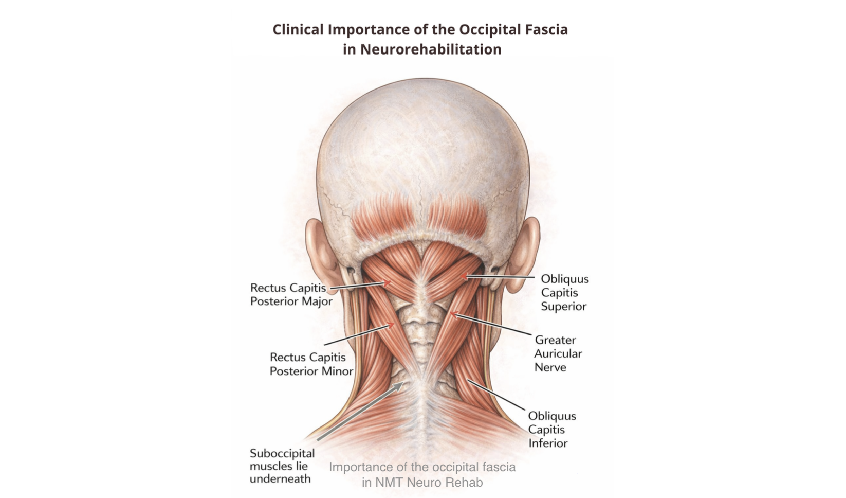
Occipital Fascia in Neurorehabilitation – Part 1
Occipital fascia in neurorehabilitation has emerged as an important area of clinical interest because of its central role in connecting the cranial base with the cervical spine, vascular structures, neural tissues and fascial networks. As a strategic interface for circulation, lymphatic and glymphatic flow, proprioception and neuromuscular regulation, the occipital fascia influences both local tissue physiology and broader neurological function. NeuroMuscular Taping (NMT) applies decompression principles to this region to improve tissue mobility, optimize fluid dynamics and support functional recovery in neurological rehabilitation.ntroduction
The occipital region forms one of the most important anatomical junctions within the human body, linking the cranial base, upper cervical spine, vascular pathways, lymphatic networks and neural structures. Occipital fascia in neurorehabilitation is increasingly recognized as a critical therapeutic target because changes in fascial tension, tissue pressure and mobility can influence circulation, cerebrospinal fluid dynamics, lymphatic drainage and neuromuscular control. These interconnected physiological mechanisms play a significant role in both normal neurological function and the recovery process following neurological injury or disease.
Furthermore, the occipital fascia should not be viewed simply as a passive connective tissue layer. Instead, it functions as a dynamic pressure-regulating interface that continuously responds to movement, muscle activity and fluid exchange. Through its decompression methodology, NeuroMuscular Taping (NMT) seeks to restore physiological pressure gradients, improve microcirculation, facilitate lymphatic and glymphatic flow, and enhance sensory input from the cranio-cervical region. By integrating biomechanical, vascular and neurophysiological principles, this approach provides clinicians with an evidence-informed strategy for supporting neuroplasticity, motor control and functional recovery across a wide range of neurological conditions.
A Strategic Interface for Integrating NeuroMuscular Taping in Circulation, Fluid Dynamics, and Neurorehabilitation
The occipital region represents a critical anatomical and biomechanical junction between the cranial base and the upper cervical spine. Deep to the superficial tissues lies the occipital fascia, a dense multilayered connective system integrating musculoskeletal, vascular, neural, and lymphatic structures. Owing to its anatomical continuity and biomechanical role, alterations in fascial tension or regional compression can significantly influence local hemodynamics, interstitial fluid transport, tissue pressure gradients, and neuromuscular regulation.
Within neurological and neurorehabilitation practice, the occipital fascial complex should therefore be regarded as a primary therapeutic target. Its strategic location allows local mechanical interventions to exert system-wide physiological and functional effects, particularly when addressed through decompressive therapeutic strategies such as NeuroMuscular Taping (NMT).
The Occipital Region as a Pressure-Sensitive Interface
The occipital fascia is positioned at the cranio-cervical junction where muscle tone regulation, connective tissue mechanics, vascular flow, cerebrospinal and interstitial fluid dynamics, and neural control mechanisms converge. This region functions as a dynamic pressure-modulating interface rather than a passive structural layer that is continuously regulated through body movement.
Physiological function depends on the mechanical environment generated by surrounding tissues, especially the suboccipital musculature. Balanced muscle tone and fluid cranio-cervical mobility help maintain:
- Adequate interstitial space
- Optimal fascial glide
- Efficient venous and lymphatic transit
- Stable neuromechanical coupling
Conversely, muscular hypertonicity, immobility due to central nervous system pathological conditions and segmental stiffness promote fascial densification, disturb local pressure gradients, and impair microcirculatory and fluid exchange processes.
The Occipital Fascia as a Pressure-Regulating Interface
Hypertonicity and Fascial Compression. Persistent contraction of suboccipital and deep cervical musculature imposes sustained mechanical loading across fascial planes, leading to:
- Increased regional tissue density
- Reduced fascial elasticity
- Narrowing of inter-fascial sliding interfaces
- Elevated interstitial pressure
This compressive state limits fascial deformation capacity and elastic recoil—properties essential for physiological load distribution, tissue adaptability, and fluid regulation.
Restoration of Tissue Compliance. Normalization of muscle tone restores the biomechanical behavior of fascial tissues:
- Re-establishment of mobility between fascial layers
- Reduction in tissue viscosity
- Improved elastic recoil
- Resumption of micro-sliding between connective interfaces
These changes create the mechanical preconditions necessary for physiological pressure modulation within fascial compartments.
Local Negative Pressure as a Core Physiological Mechanism
Healthy fascial systems function as dynamic biomechanical regulators rather than passive connective envelopes. Their structural elasticity, multilayered organization, and viscoelastic behavior allow continuous modulation of pressure within interstitial and microvascular environments. A central feature of this regulation is the generation of subtle cyclical pressure oscillations driven by tissue elasticity and physiological movement.
Definition and Physiological Significance. Local negative pressure refers to a relative reduction in subcutaneous and interstitial pressure that secondarily lowers resistance within capillary and lymphatic vessels, thereby facilitating:
- Microvascular perfusion
- Venous return
- Lymphatic uptake
- Interstitial fluid exchange
- Fascia gliding
Local negative pressure does not represent literal suction or vacuum formation. Instead, it refers to:
“A transient reduction in interstitial hydrostatic pressure relative to adjacent tissue compartments that increases the functional surface area of the microcirculation and lymphatic systems. The increase in negative pressure is continuously modified during body movement.”
This pressure differential establishes favorable gradients that facilitate fluid displacement and microvascular exchange.
Circulation and Fluid Dynamics at the Cranio-Cervical Junction
a) Microvascular Perfusion. Reduced interstitial pressure lowers extramural compression on capillaries, allowing: • Increased capillary lumen diameter • Recruitment of previously collapsed capillaries • Expanded surface area for exchange. This enhances oxygen delivery and metabolic waste removal.
b) Venous Return. Venous circulation is pressure-dependent and benefits from reduced surrounding tissue resistance which: • Lower external pressure facilitates venous filling • Improve compliance of venous walls • Enhance cranio-caudal blood return.
c) Lymphatic Uptake. Initial lymphatic vessels rely on pressure gradients for fluid entry that: • Decrease interstitial pressure opens anchoring filaments • Inter-endothelial gaps widen • Protein-rich interstitial fluid enters lymphatic capillaries. This supports edema control and inflammatory resolution.
d) Interstitial Fluid Exchange. Negative pressure enhances bidirectional exchange governed by Starling forces that: • Improve filtration and reabsorption • Enhance osmotic flux • Maintain of tissue hydration • Optimize nutrient delivery.
Capillary recruitment expands the exchange interface for transcapillary fluid movement, enhancing filtration and reabsorption processes according to Starling forces. In combination with osmotic gradients, this supports tissue hydration, metabolic exchange, and nutrient delivery—particularly in metabolically active or inflamed tissues.
Pressure Oscillations and the Role of the Glymphatic and Lymphatic Systems
Fascia is composed of collagenous and elastin fibers embedded within a hydrated extracellular matrix rich in glycosaminoglycans and proteoglycans. This composition provides: • Elastic recoil capacity • Viscoelastic deformation under load • Reversible shape adaptation • Mechanical energy storage and release.
During normal movement, postural adjustments, respiration, and muscle activity, fascial layers undergo rhythmic deformation. Each cycle consists of:
- Compression Phase • Temporary reduction of interstitial space • Mild increase in local tissue pressure • Mechanical support for venous propulsion
- Recoil / Decompression Phase • Elastic restoration of tissue length • Expansion of interstitial compartments • Reduction in local pressure
- Fascial Mobility as a Prerequisite. For pressure oscillations to occur efficiently, fascia must retain: ✔ Layered glide capacity ✔ Elastic recoil potential ✔ Hydrated extracellular matrix ✔ Freedom from densification
It is during the decompressive phase of tissue recoil that a relative reduction in local interstitial pressure is generated. As fascial layers elastically return to their resting configuration, subtle expansion of the extracellular space occurs, creating a transient low-pressure environment that favors fluid movement and microvascular exchange. When fascial tissues lose their mobility—due to densification, adhesions, or sustained muscular hypertonicity—this natural oscillatory behavior is attenuated. The resulting reduction in pressure variability impairs fluid dynamics, promoting interstitial stagnation, limiting nutrient delivery, and compromising cellular metabolic processes.
From a clinical perspective, effective modulation of tissue pressure supports multiple physiological functions. Normalization of interstitial dynamics helps reduce local fluid congestion and improves oxygen diffusion to metabolically active tissues. Enhanced microcirculatory exchange facilitates more efficient nutrient transport and waste removal, contributing to improved metabolic performance. Simultaneously, reduction in mechanical stress on nociceptive structures decreases pain sensitivity, while improved tissue compliance and fluid balance support more coordinated and energy-efficient neuromuscular activity.
These mechanisms assume particular importance in pressure-sensitive anatomical regions such as the cranio-cervical junction, where small alterations in fascial mechanics can influence vascular flow, neural regulation, and postural control. Optimizing local pressure regulation in this region therefore represents a key therapeutic objective in circulatory management and neurological rehabilitation.
NeuroMuscular Taping and Fascial Decompression
NeuroMuscular Taping (NMT), developed in 2003, is founded on the clinical principle of cutaneous and fascial decompression achieved through a tape application without external tension. The methodology promotes tissue lifting, space creation, and normalization of pressure within superficial and deep compartments, facilitating physiological fluid dynamics and neuromuscular regulation.
Mechanisms by Which NMT Facilitates Negative Pressure
- Mechanical Decompression. The decompressive configuration of NMT reduces external compressive forces on soft tissues, permitting subtle volumetric expansion.
- Fascial Recoil. Elastic connective fibers undergo assisted recoil through eccentric elongation forces induced by tape–skin interaction, increasing interstitial and muscular volume.
- Pressure Gradient Formation. Incremental tissue expansion lowers local interstitial pressure relative to adjacent compartments. Continuous and graded decompression—modulated by physiological movement—maintains favorable pressure differentials.
- Fluid Mobilization. Biological fluids move from regions of higher resistance toward areas of lower resistance. By reducing interstitial impedance, NMT supports vascular efficiency and lymphatic drainage at the cranial base.
NMT Improves Cranio-Cervical Mobility as a Fascial Pump Mechanism
Movement-Induced Pressure Cycling is a gentle cranio-cervical motion that produces cyclical compression–decompression phases that:
- Generate rhythmic pressure oscillations
- Promote interstitial fluid displacement
- Assist venous and lymphatic return
- Support tissue perfusion with minimal muscular effort
This mechanism functions as decompression taping enhances a biomechanical pump supporting regional fluid physiology.
Consequences of Restricted Mobility creates movement restriction and protective muscle guarding:
- Attenuate pressure oscillations
- Reduce tissue hydration dynamics
- Promote interstitial stagnation
- Increase nociceptive mechanosensitivity
Thus, mobility is fundamental not only for joint kinematics but also for tissue fluid regulation.
Functional Outcomes of Optimized Occipital Mechanics Through NeuroMuscular Taping Treatments
When NeuroMuscular Taping (NMT) is applied using decompressive principles to the occipital and cranio-cervical region, it facilitates restoration of physiological muscle tone and promotes fluid, coordinated head–neck mobility. By gently lifting the skin and underlying fascial layers without external tension, NMT reduces compressive loading within the suboccipital compartment and creates space within superficial and deep connective tissues. This decompressive effect enables fascial planes to recover their natural capacity for glide and adaptive motion, improving the distribution of mechanical forces across the cranio-cervical junction.
As tissue compliance improves, interstitial pressure progressively normalizes, establishing a more favorable microenvironment for vascular and fluid exchange. Reduced external resistance on low-pressure venous structures enhances cranial venous outflow, while improved pressure gradients facilitate lymphatic uptake and drainage. These changes support more efficient clearance of interstitial fluids and metabolic by-products, contributing to improved tissue perfusion and regional homeostasis.
In parallel, decompression of mechanosensitive neural interfaces reduces nociceptive input and supports neuromodulation of cervical muscle tone. With diminished tissue resistance and improved fascial elasticity, cranio-cervical movements become smoother, more coordinated, and biomechanically efficient. Through these combined mechanisms, NMT-assisted optimization of occipital mechanics enhances postural control, sensorimotor integration, and functional performance—key therapeutic objectives in musculoskeletal and neurological rehabilitation.
Clinical Relevance in NeuroMuscular Taping–Based Neurorehabilitation
Neurological disorders commonly alter the biomechanical and neurophysiological balance of the cranio-cervical region. Patients frequently present with axial rigidity, persistent hypertonic extensor patterns, cranio-cervical compression, and reduced capacity for adaptive motor control. These features are particularly evident in conditions such as stroke, Parkinsonian syndromes, cerebral palsy, traumatic brain injury, and spinal cord pathologies, where abnormal tone regulation and impaired postural reflexes disrupt the functional relationship between the head, neck, and trunk. The occipital region becomes a zone of mechanical overload, fascial densification, and altered sensory input, further aggravating motor inefficiency and postural instability.
Continuous 24/7 application of NeuroMuscular Taping (NMT) using decompressive principles provides a sustained mechanical and sensory intervention that directly addresses these dysfunctions. By gently lifting the skin and underlying fascial layers without external tension, NMT promotes the creation of a decompressive tissue environment at the cranio-cervical junction. This space-restoring effect reduces compressive stress on myofascial, neurovascular, and connective structures, facilitating improved venous outflow and lymphatic drainage while enhancing interstitial fluid exchange. The resulting improvement in tissue perfusion supports metabolic recovery and reduces local inflammatory and nociceptive drivers that often perpetuate abnormal tone.
From a neuromotor perspective, the continuous cutaneous stimulation provided by NMT contributes to modulation of mechanoreceptor activity and afferent sensory input. This sustained sensory feedback assists in recalibrating sensorimotor integration pathways that are frequently disrupted in central nervous system disorders. Improved proprioceptive signaling from the cranio-cervical region supports more efficient head–neck alignment, facilitates normalization of extensor–flexor muscle balance, and enhances the coordination of postural reflex strategies.
As cranio-cervical mechanics become more efficient, patients demonstrate improved head control, smoother transitional movements, and better distribution of axial loads across the spine. Enhanced postural stability reduces compensatory muscular overactivity and promotes more energy-efficient movement patterns during functional tasks such as sitting, standing, gait initiation, and upper-limb activity. These changes are particularly relevant in neurorehabilitation, where restoring adaptive motor control and postural organization is central to functional recovery.
Accordingly, occipital decompression should be regarded as a foundational component of comprehensive neurological rehabilitation protocols employing NeuroMuscular Taping. Addressing the cranio-cervical junction through sustained decompressive taping not only improves local tissue mechanics but also exerts system-wide effects on circulation, sensorimotor regulation, and postural control—key determinants of rehabilitation outcomes in patients with central and spinal neurological conditions. Specific NMT treatments and pathology protocols will be outlined in Part 2.
References
- Andi WS, Wahida W, Blow D. Application of neuromuscular taping on occipital neuralgia: Case study. October 2019. Journal of the Neurological Sciences 405:46-47.https://www.sciencedirect.com/science/article/pii/S0022510X19321148
- Blow D. NeuroMuscular Taping: From Theory to Practice. (2012). ISBN 10: 1467530360 / ISBN 13: 9781467530361
- Blow D. Neuromuscular Taping: Treatment of Edemas, Hematoma. (2013) ISBN 10: 8870515990 / ISBN 13: 9788870515992
- Costantino C, Pedrini MF, Licari O. Neuromuscular taping versus sham therapy on muscular strength and motor performance in multiple sclerosis patients. Disabil Rehabil. 2016;38(3):277–281. https://doi.org/10.3109/09638288.2015.1038365
- Findley TW. Fascia research from a clinician/scientist’s perspective. Int J Ther Massage Bodywork. 2009;2(4):1–6. https://doi.org/10.3822/ijtmb.v2i4.63
- Kristianto H, Ahsan A, Mukaroma L, Akhsinnadya F, Indri MC, Damayanti N, Kusbandiyah I, Rohyani RT, Blow D, Rias YA, Wibisono AH, Noviyanti LW, Nasution TH. Effects of neuromuscular taping form I on wound temperature and erythema in diabetic foot ulcer: a preliminary study. J Wound Care. 2024;33(6):425–430. https://doi.org/10.12968/jowc.2020.0291
- Levick JR, Michel CC. Microvascular fluid exchange and the revised Starling principle. Cardiovasc Res. 2010;87(2):198–210. https://doi.org/10.1093/cvr/cvq062
- Maggi L, Celletti C, Mazzarini M, Blow D, Camerota F. Neuromuscular taping for chronic non-specific low back pain: a randomized single-blind controlled trial. Aging Clin Exp Res. 2021. https://doi.org/10.1007/s40520-021-02029-0
- Mortimer PS, Rockson SG. New developments in lymphatic disease. J Clin Invest. 2014;124(3):915–921. https://doi.org/10.1172/JCI71608
- Schleip R, Müller DG. Training principles for fascial connective tissues. J Bodyw Mov Ther. 2013;17(1):103–115. https://doi.org/10.1016/j.jbmt.2012.06.007
- Shah M, Julu POO, Monro JA, Coutinho J, Ijeh C, Puri BK. Neuromuscular taping reduces blood pressure in systemic arterial hypertension. Med Hypotheses. 2018;116:30–32. https://doi.org/10.1016/j.mehy.2018.04.014
- Standring S, ed. Gray’s Anatomy: The Anatomical Basis of Clinical Practice. 42nd ed. Elsevier; 2020.
- Stecco A, Stern R, Fantoni I, De Caro R, Stecco C. Fascial disorders: implications for treatment. Surg Radiol Anat.2016;38(7):787–794. https://doi.org/10.1007/s00276-016-1652-8
- Stecco C, Macchi V, Porzionato A, Duparc F, De Caro R. The fascia: the forgotten structure. J Bodyw Mov Ther.2011;15(2):127–138. https://doi.org/10.1016/j.jbmt.2010.11.010
- Swartz MA. The physiology of the lymphatic system. Adv Drug Deliv Rev. 2001;50(1–2):3–20. https://doi.org/10.1016/S0169-409X(01)00150-8
- Wilke J, Krause F, Vogt L, Banzer W. What is evidence-based about myofascial chains? Arch Phys Med Rehabil.2016;97(3):454–461. https://doi.org/10.1016/j.apmr.2015.07.023
- Yahia LH, Rhalmi S, Newman N, Isler M. Sensory innervation of human thoracolumbar fascia. Spine (Phila Pa 1976).1992;17(11):1313–1317. https://doi.org/10.1097/00007632-199211000-00006
See the LINKEDIN NMT Newsletter

Are you interested in enrolling in an NMT Course?
The NeuroMuscular Taping Institute provides certification courses in physical rehabilitation, neurology, oncology, post surgical rehab, occupational therapy, speech therapy, remedial therapy, nursing as well vascular and lymphedema treatment and management. All trainings are classroom settings to enhance skill development and clinical reasoning creating optimum therapeutic results. ONLINE courses are not available. Please contact your local course provider or visit the NMT WEB SITE for course programs and dates.
For more details, course listings, and registration for an upcoming course CLICK HERE
Do you require validation of your NMT methodology for research purposes?
A detailed Neuromuscular Taping methodology is crucial in any research project for several reasons. Repeatability, Transparency, Validity and Reliability of your treatment choices will not only have an impact on your research but also an impact on future therapeutic choices. For more detailed information about the NMT, please contact us at: david.blow@nmtinstitute.org
Know someone who might be interested in this newsletter? Share it with them.